Cancer
Cancer Research
Positron Emission Tomography (PET) imaging is regularly used to aid diagnosis of several different types of cancer.
A fluorine-containing version of glucose, Fluorodeoxyglucose or FDG, is the radioactive tracer most commonly used. A 'tracer' is a radioactive substance given in extremely small quantities, so that it should not have any chemical or biochemical effect. The technique is sometimes called FDG-PET to distinguish it from PET imaging with other tracers.
FDG is a useful tracer because tumour cells often use glucose for energy metabolism more actively than normal cells. After injection of FDG, the patient is allowed to rest quietly for one hour before PET imaging. The images then help to show whether primary or metastatic tumours are present.
In some cases, FDG-PET may also be useful in evaluating whether a cancer treatment is working or in helping to define the size and position of the tumour, so that radiotherapy can hit the required target and minimise damage to normal tissues.
For research purposes, other PET tracers are being developed. These can take advantage of other disturbed features of tumour cell metabolism:
- Fluorocholine (FCho) is used in prostate cancer
- Fluorethyltyrosine (FET) can be used to monitor brain tumours
- Radioactive versions of new drugs may be used to test how well the drug is taken into cancer cells
In addition to fluorine (18F) based PET tracers, some radioactive metals have become important in cancer diagnosis and research:
- Gallium (68Ga) can be attached to probes specific for prostate cancer, making them especially useful in identifying tumour metastases
- Zirconium (as 89Zr) has a much longer half-life than typical PET isotopes. It can be attached to antibodies (which typically take a few days to localise to target tumour cells) so that their relatively slow accumulation can be followed
The NUTH Cancer Research Centre has active trial programmes running which could benefit from access to whole body PET. Dr Rachel Pearson and Bhavan Rai are interested in imaging and treating prostate and renal cancers using FDG, 18F-PSMA, and 89Zr-tagged antibodies.
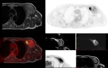
SarcoPET Study
We carried out a small feasibility study using FDG-PETMR for staging of Myxofibrosaroma.
Myxofibrosarcomas (MFS) are soft tissue tumours that can develop both centrally and in the limbs. The optimal treatment includes surgery, but this is often compromised as MFS can infiltrate or blend into the surrounding normal tissues making it difficult to distinguish between normal tissue and tumour at surgery.
We concluded that both FDG-PET and DWI MRI potentially offer more accurate local staging of myxofibrosarcomas and could reduce re-excision rates and the requirement for post-operative radiotherapy.
Hazel McCallum is developing the use of PET-MRI for radiotherapy planning
JJ Wyatt has designed a project to use FDG PET-MRI to stage rectal cancers.
In collaboration with Dr. Satomi Miwa in Newcastle, we are studyiing PET probes for senescent cells, based on galactose (18F-PyGal is an analogue of galactose that is trapped in senescent cells because of changes in an enzyme that metabolises galactose). Although these 'sleeping' cells are of importance in ageing research, they are also relevant in cancer research because both radiotherapy and chemotherapy can result in cells becoming senescent. We plan to study senescence of tumour cells before and after therapy.
Further information
Find out more about our research in this area:
- clinical trials
- radiotherapy planning
-
PET imaging is widely used in cancer diagnosis and management as described on the Cancer Research UK website,
https://www.cancerresearchuk.org/about-cancer/tests-and-scans/pet-scan - where specific information about PET-MRI is also provided. https://www.cancerresearchuk.org/about-cancer/tests-and-scans/pet-mri-scan