Multi-electrode Array (MEA) Facility
Equipment
Microscope: Nikon FN1 with BsiExpress eqipped with :
- Faraday cage
- Manual Stage
- Temp. control
- BioCam Duplex (MEA)
- Polygon 1000 (DMD)
- Peristaltic and fast switching perfusion systems
- CoolLed pE800 and 340 Fura
")
Array Platform
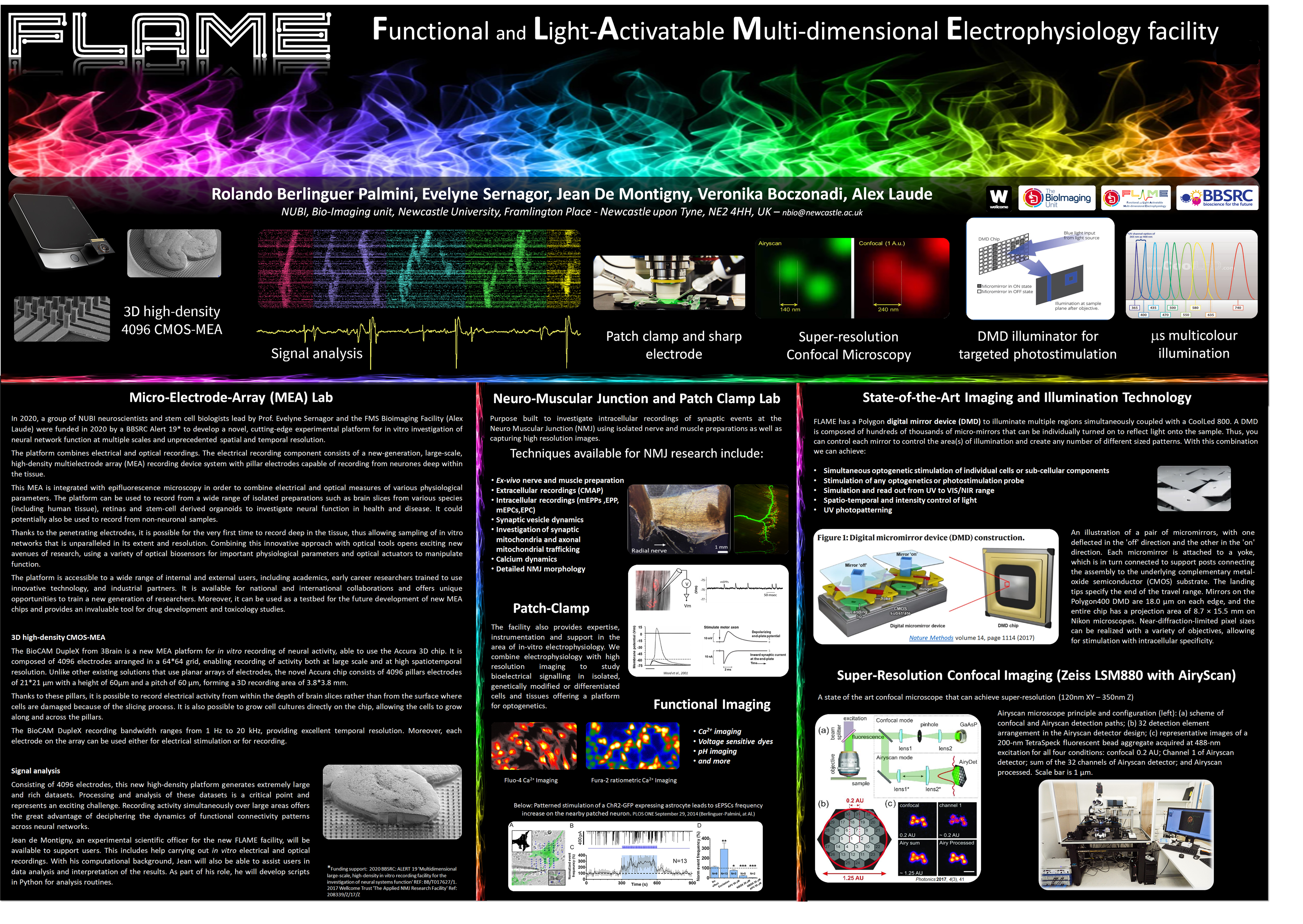
3D high-density CMOS-MEA
The BioCAM DupleX from 3Brain is a new MEA platform for in vitro recording of neural activity, able to use the Accura 3D chip. It is composed of 4096 electrodes arranged in a 64*64 grid, enabling recording of activity both at large scale and at high spatiotemporal resolution. Unlike other existing solutions that use planar arrays of electrodes, the novel Accura chip consists of 4096 pillars electrodes of 21*21μm with a height of 60μm and a pitch of 60μm, forming a 3D recording area of 3.8*3.8 mm.Thanks to these pillars, it is possible to record electrical activity from within the depth of brain slices rather than from the surface where cells are damaged because of the slicing process. It is also possible to grow cell cultures directly on the chip, allowing the cells to grow along and across the pillars. The BioCAM DupleX recording bandwidth ranges from 1Hz to 20kHz, providing excellent temporal resolution. Moreover, each electrode on the array can be used either for electrical stimulation or for recording.
Analysing electrophysiological data
Signal analysis
Consisting of 4096 electrodes, this new high-density platform generates extremely large and rich datasets. Processing and analysis of these datasets is a critical point and represents an exciting challenge. Recording activity simultaneously over large areas offers the great advantage of deciphering the dynamics of functional connectivity patterns across neural networks. Jean de Montigny, an experimental scientific officer for the new FLAME facility, will be available to support users. This includes help carrying out in vitro electrical and optical recordings. With his computational background, Jean will also be able to assist users in data analysis and interpretation of the results. As part of his role, he will develop scripts in Python for analysis routines.


Location
Address:Cookson BuildingGround floorRoom G31
Dental School
The Medical School
Framlington Place
Newcastle Upon Tyne
NE2 4HH
UK
MEA Gallery
Mouse Retina Spontaneous waves. - Credit for the processing to Dr. Michael Savage (Prof. Sernagor Lab) and for the Calcium images to Dr Jean De Montigny.
Organotipic slices. - Credit for the sample preparation to Dr. Faye McLeod and for the Calcium imagesto Dr Jean De Montigny.