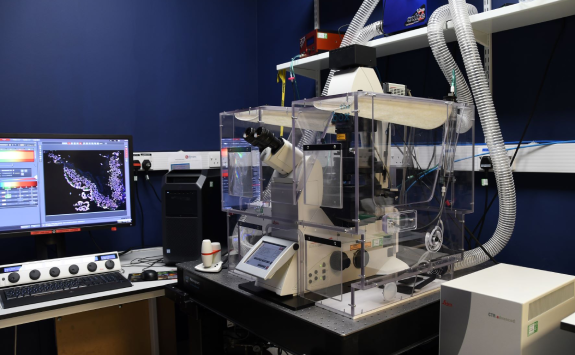
Leica SP8 DLS
Modalities: invert, confocal, live cell, light sheet
The SP8 is a point scanning confocal microscope based around a fully motorised, inverted DMi8 body with hardware-enabled focus correction. The system has two photomultipliers and two HyD detectors for fluorescence imaging and one for transmitted light detection.
The HyD detectors are more sensitive than conventional PMTs, allowing imaging of very weak fluorescence, and can also be used in photon counting mode, providing truly quantitative imaging. Incorporated into the confocal scan-head are both resonant (fast scanning) and non-resonant (conventional scanning) scanning mirrors. This allows experimenters to have the option of imaging in high resolution (up to 8192 x 8192 pixels) or at high speed (290fps at 512 x 16 pixels). Resonant scanning mode has added benefits; for live-cell experiments, photo-sensitive specimens remain alive for longer and fluorophore bleaching is significantly reduced.
The system will also perform regular 2D, 3D and 4D imaging.
The microscope is fully-enclosed in a heated environmental chamber and has the ability to set and maintain temperature and CO2.
For larger specimens, the confocal can be utilised as a Digital Light Sheet system (DLS), allowing very fast multicolour z stack imaging through samples embedding in aqueous medium, ideal for fly and fish embryo or organoid imaging studies.
Specification
Objectives
Magnification |
NA |
Coverslip |
Other modes |
Immersion |
|---|---|---|---|---|
For DLS | ||||
| 2.5 | 0.07 | excitation objective | Air | |
| 10 | 0.3 | emission objective | Water | |
| 25 | 0.95 | emission objective | Water | |
For Confocal | ||||
| 5 | 0.15 | Air | ||
| 10 | 0.4 | Air | ||
| 20 | 0.75 | 0.17 | DIC | Air |
| 40 | 0.55 | 0.17 | Water | |
| 40 | 0.85 | 0.17 | Air | |
| 40 | 1.3 | 0.17 | Oil | |
| 63 | 1.4 | 0.17 | Oil |
Filter (epi)
Cube |
Example fluorescence |
Excitation |
Dichroic |
Emission |
Part number |
|---|---|---|---|---|---|
| Leica DAPI | DAPI, Hoechst | 340-380 | 400 | 420-(LP) | |
| Leica FITC | GFP, FITC, AF488 | 450-490 | 510 | 515 (LP) | |
| Leica RHOD | mCherry, AF594 | 541-551 | 580 | 565-605 |
Lasers Lines
- Diode 405nm, solid state 488nm, 552nm, 638nm
Detectors
- 2 x HyD (hybrid detectors) for fluorescence or photon counting
- 2 x PMT for fluorescence
- 1x PMT for transmission