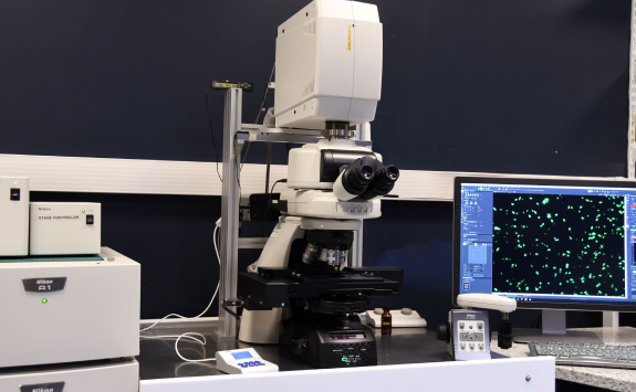
Nikon A1
Modalities: upright, confocal
Overview
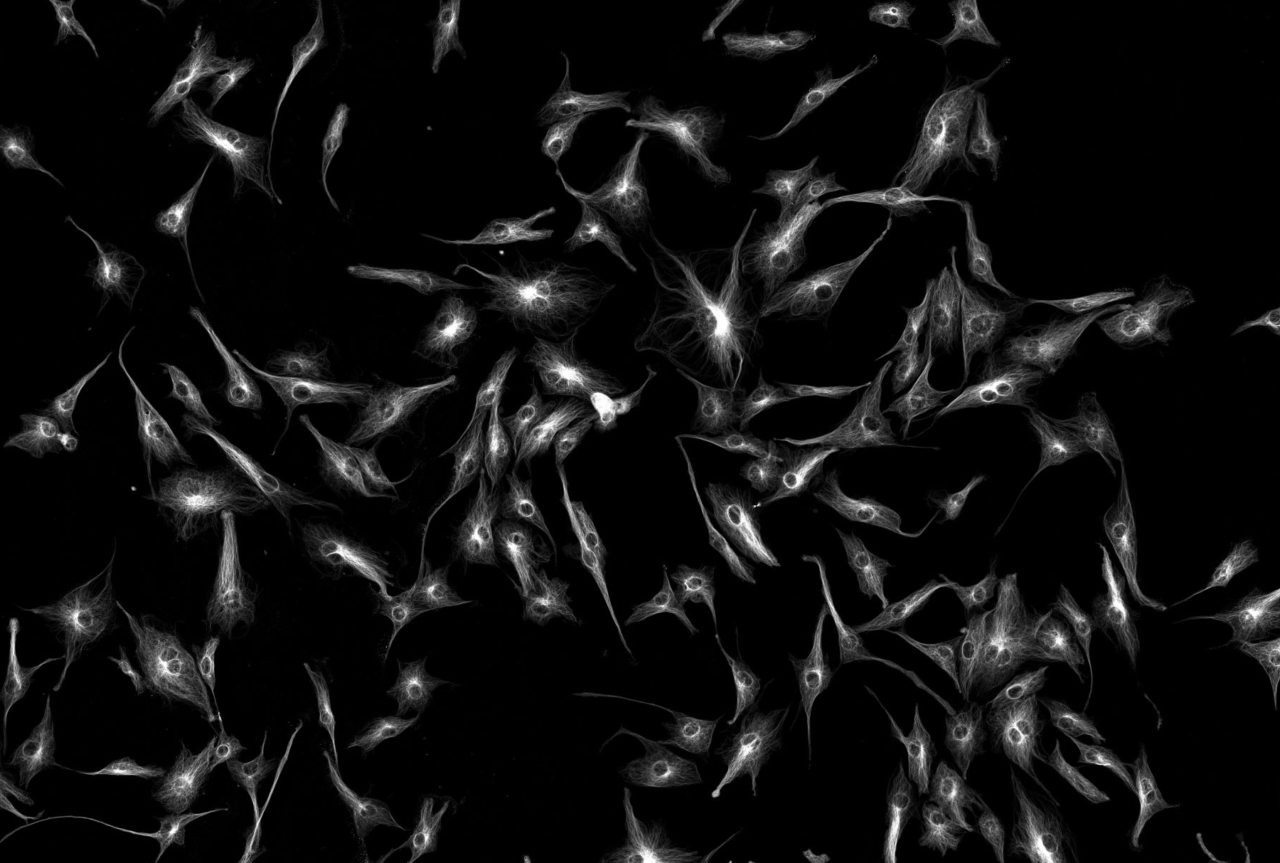
The Nikon A1+ is a point scanning confocal microscope based around a fully motorized, upright Nikon Ni body.
The system has four photomultipliers (two of which are GaAsP) for fluorescence imaging and an additional one for transmitted light detection.
Imaging can be performed at up to 4096 x 4096 pixel resolution.
The system is run through Nikon’s versatile Elements software package and incorporates wizards to perform common imaging tasks such as:
- FRET
- colocalisation
- data quantification
Imaging modes
- Multi point x,y (2D) with tiling
- Volume x,y,z (3D)
- Time lapse x,y,z,t (4D)
Specification
Here you can find the specifications for this microscope.
Objectives
Magnification |
NA |
Coverslip |
Other modes |
Immersion |
|---|---|---|---|---|
| 4 | 0.2 | Air | ||
| 10 | 0.45 | 0.17 | Air | |
| 20 | 0.75 | 0.17 | Air | |
| 40 | 0.95 | 0.17 | Air | |
| 40 | 1.3 | 0.17 | Oil | |
| 60 | 1.4 | 0.17 | Oil |
Filters (epi)
Cube |
Example Fluorophores |
Excitation |
Dichroic |
Emission |
Part Number |
|---|---|---|---|---|---|
| Chroma 49000-ET | DAPI, Hoechst | 325-375 | 400 | 435-485 | |
| Chroma 49002-ET | GFP, FITC, AF488 | 450-490 | 495 | 500-550 | |
| Chroma 49008-ET | mCherry, AF594 | 540-580 | 585 | 592-662 |
Light Source (epi)
CoolLed pE 300 (370, 460, 550)
Laser Lines
405nm, 488nm, 561nm, 647nm
Detectors (cameras or PMTs)
2 PMT, 2 GaAsP