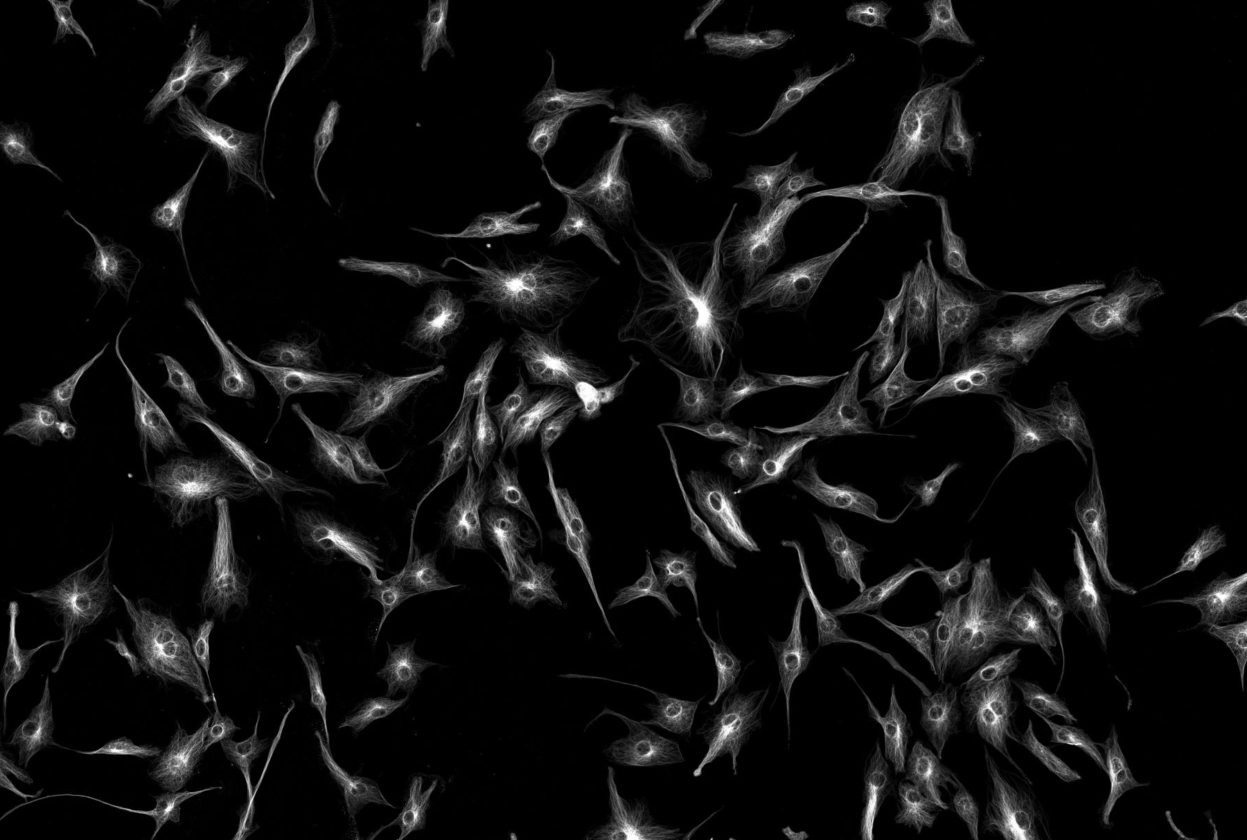
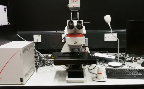
Leica DM6
Modalities: upright, widefield, stereology
Overview
This microscope is a fully automated widefield system for fluorescence and brightfield imaging.
The DM6 has a full range of objectives (10- 100x), monochrome and color cameras and filter sets to enable fluorescence imaging of up to 5 distinct channels from UV to far red excitation.
Stage and z are also motorized, allowing multichannel z stacks in multiple positions or tile-scans.
The system can be operated using one of two software package; Leica LASX or MBF StereoInvestigator.
Specification
Here you can find the specification for this microscope.
Objectives
Magnification |
NA |
Coverslips |
Other Modes |
Immersion |
|---|---|---|---|---|
| 2.5 | 0.07 | Air | ||
| 5 | 0.15 | Air | ||
| 10 | 0.4 | 0.17 | Air | |
| 20 | 0.7 | 0.17 | Ph 2 DIC | Air |
| 40 | 0.85 | 0.17 | Air | |
| 63 | 1.32 | 0.17 | Ph3 CS | Oil |
| 100 | 1.44 | 0.17 | DIC | Oil |
Filters (epi)
Cube |
Example Fluorophores |
Excitation |
Dichroic |
Emission |
Part Number |
|---|---|---|---|---|---|
| Leica DAPI | DAPI, Hoechst | 325-375 | 400 | 435-485 | |
| Leica L5 | GFP, FITC,AF488 | 460-500 | 505 | 512-542 | |
| Leica N3 | AF555 | 540-552 | 565 | 580-620 | |
| Leica TXR | AF568, AF594 | 540-580 | 585 | 593-667 | |
| Leica Y5 | AF647 | 608-648 | 669 | 672-712 |
Light source (epi)
Sola Light SE - Broad spectrum fluorescent light
Cameras
Hamamatsu Flash 4, Monochrome (for fluorescence imaging)
Leica DFC450C, Colour (for brightfield HC)

