Impact and Outreach
Take a closer look at research highlights, achievements, engagement, awards and impact in 2020-2021 enabled by our facility.
Awards
Huygens Image Contest 2025
Congratulations to Dr. Ana Andjelković and Dr. Rolando Berlinguer Palmini, Faculty of Medical Sciences and BioImaging Unit, Newcastle University, UK for first prize.
This year's winner was selected because it combines a wonderfully difficult feat of microscopy with Huygens' powerful restoration, and is given form in an effective visualization. The movie shows live-cell STED microscopy of localization and quantity of mitochondrial DNA (mtDNA) under conditions of perturbed mitochondrial gene expression in the SH-SY5Y cell line, the aim being to capture these structures at high spatial and temporal detail, while minimally affecting live cells. The captured images were deconvolved with Huygens, realigned for Z and XY drift if needed and rendered with the Huygens Surface Renderer.
Huygens Image Contest 2021
Congratulations to Dr. Rolando Berlinguer Palmini (BioImaging Unit) and Dr. Christin Albus (Mitochondrial Research Group), Newcastle University, UK for first prize.
Christmas Lights & Christmas Tree - Challenging four colour STED 3D stack combined with RNA FISH! Leica STED image was deconvolved and visualized with Huygens Professional! U2OS cells with mitochondrial outer membrane Tom20 (AF488 - White), ND1 mRNA FISH Quasar 570 ND1 NADH dehydrogenase subunit 1 (Green), ND2 mRNA FISH CalFluor 610 NADH dehydrogenase two (ND2) protein (Red), mitochondrial RNA-binding protein GRSF1 (RNA granule) - Abberior star red (Blue). Tom20 is MIP rendered (white) and other channels are surfaces rendered with Huygens.
Huygens Image Contest 2020
In the 2020 Huygens Image Contest, Dr. Rolando Berlinguer Palmini (BioImaging Unit) and Dr. Matthew Zorkau (Mitochondria Research Group), at Newcastle University received the third Prize for their image.
This has proven to be a crucial image to show the submitochondrial location of newly synthesised proteins. The green rendered fluorescent signal represents a newly synthesised mitochondrial protein and the red is an immunofluorescent antibody to a mitochondrial protein found in the inner boundary membrane of the mitochondrion (TIM23). This Huygens deconvolved image visualized with Huygens Surface Renderer shows a section of the human mitochondrial network taken at STED nanoscopy resolution using AF594 depleted with a 775nm STED laser and AF532 depleted with a 660 nm STED laser. You can read about this more on the Huygens website.
Front covers
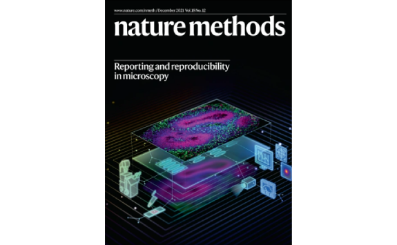
Cover of Nature Methods
This Focus issue features a suite of papers offering guidelines and solutions for collecting and reporting microscopy metadata. Papers come from the Quality Assessment and Reproducibility for Instruments & Images in Light Microscopy (QUAREP-LiMi) group and colleagues. The Bio-imaging Unit are a part of this. We are delighted that our work is featured and we claimed the cover for this issue. For more information visit our QUAREP site and read more on the Nature Methods website.
Nature Cancer Cover
Congratulations to all authors especially Anna Smith, Julia Whitehall, Laura Graves and all the other authors for their paper which published an immunofluorescent image that made the cover of Nature Cancer. The winning image shows co-labelling of OXPHOS proteins and SSP enzymes in normal human colonic epithelium. Cut sections were imaged using Zeiss LSM800 confocal microscope and were analysed using ImageJ software (NIH).
The full article can be found on Nature Cancer.
Abstract: Oxidative phosphorylation (OXPHOS) defects caused by somatic mitochondrial DNA mutations increase with age in human colorectal epithelium and are prevalent in colorectal tumors, but whether they actively contribute to tumorigenesis remains unknown. Here we demonstrate that mitochondrial DNA mutations causing OXPHOS defects are enriched during the human adenoma/carcinoma sequence, suggesting that they may confer a metabolic advantage. To test this, we deleted the tumor suppressor Apc in OXPHOS-deficient intestinal stem cells in mice. The resulting tumors were larger than in control mice due to accelerated cell proliferation and reduced apoptosis. We show that both normal crypts and tumors undergo metabolic remodelling in response to OXPHOS deficiency by upregulating the de novo serine synthesis pathway. Moreover, normal human colonic crypts upregulate the serine synthesis pathway in response to OXPHOS deficiency before tumorigenesis. Our data show that age-associated OXPHOS deficiency causes metabolic remodelling that can functionally contribute to accelerated intestinal cancer development.
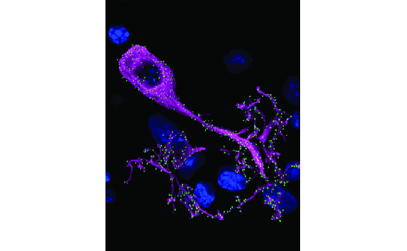
Cover of Journal of Neuroscience
Congratulations to , and
This image shows punctate neuronal nitric oxide synthase (nNOS, green) distributed on neuronal elements (magenta) in the central nucleus of the guinea pig inferior colliculus. Cell nuclei were labelled with DAPI (blue). Dual immunohistochemistry for nNOS (green) and a pan neuronal antibody mixture (PAN, magenta) show that nNOS-labeled puncta are present on the membrane of both somata and dendrites. Rendering in Imaris shows clearly how nNOS puncta (green spots) are localized on neuronal elements (magenta surfaces).
To examine the subcellular distribution of antibody labelling and the proximity of different labels, Z-stacks (Z step 0.1–0.3 μm) were acquired using a 63×oil-immersion objective on a Nikon A1+ point scanning confocal microscope with Nikon Elements software. Z-stacks were deconvolved using Huygens Essential (SVI). For more information, see the article by Olthof et al.(pages 876-887).
TV
Tyneside's Genetic Pioneers: How the NHS changed our world - BBC
Ground-breaking work by genetic and medical experts at Newcastle University and Newcastle Hospitals, housed at the Centre for Life, features in a BBC programme celebrating 70 years of the NHS.
On the anniversary, presenter, Kirsten O’Brien looks to the next 70 years and tells the story of genetic medicine on Tyneside.
All under one roof at the Centre For Life, clinicians, researchers and entrepreneurs at the Institute of Genetic Medicine and the Newcastle upon Tyne Hospitals NHS Foundation Trust are working to better understand our genetic make-up and its links to our health.
They feature in “How the NHS Changed our world: Tyneside’s Genetic Pioneers” on BBC 2 on Friday June 29 19:00.
The International Centre for Life is a science village in the heart of Newcastle. It incorporates an internationally-renowned research institute belonging to Newcastle University, several NHS clinics delivering cutting-edge fertility and regenerative medicine, and houses young and established biotechnology companies and the Life Science Centre, which is an award-winning visitor attraction and educational facility.
Images were taken on the Leica SP8 STED with Dr. Rolando Berlinguer Palmini.
How laundry is spilling plastics into the ocean - CNN
Tiny plastic particles are polluting our oceans, where they can stay for centuries, harming sea life and making their way into the human food chain. These microplastics -- fragments of plastic smaller than 5mm -- are often the result of bigger chunks being worn down by the elements. But a surprisingly large amount comes from people simply washing their clothes. You can read more about this on CNN.
Spilling plastics simulations created by Dr. Rolando Berlinguer Palmini.
Images acquired using the Nikon SMZ25 and Nikon A1R.
Publications
Publications 1 January 2017 – 1 December 2024
All publications with images acquired at the Bio-Imaging Facility: 301
- 2017: 49
- 2018: 61
- 2019: 70
- 2020: 40
- 2021: 27
- 2022: 28
- 2023: 26
Publications with acknowledgement only (specific staff member of the facility):48
Publications with staff authorship:44
Publication covers:3
Collaborations
The Bioimaging Facility's state-of-the-art equipment and services are used by internal research groups and visitors from other universities, research institutes and companies.
External users
Commercial
Academic
- Northumbria University
- Durham University
- The University of Sunderland
- The University of Edinburgh
- The University of Exeter
Outreach
We regularly participate in engagement and outreach activities and deliver themed events and small workshops and lessons to school and community groups. We also host small group University visits and can accommodate short work/day experience projects. Please access the tabs to find out more about the kind of events that we have delivered. Please contact the facility to find out more about working with, or visiting us.