Leica SP8 STED
Modalities: invert, confocal, live cell, super-resolution
Overview
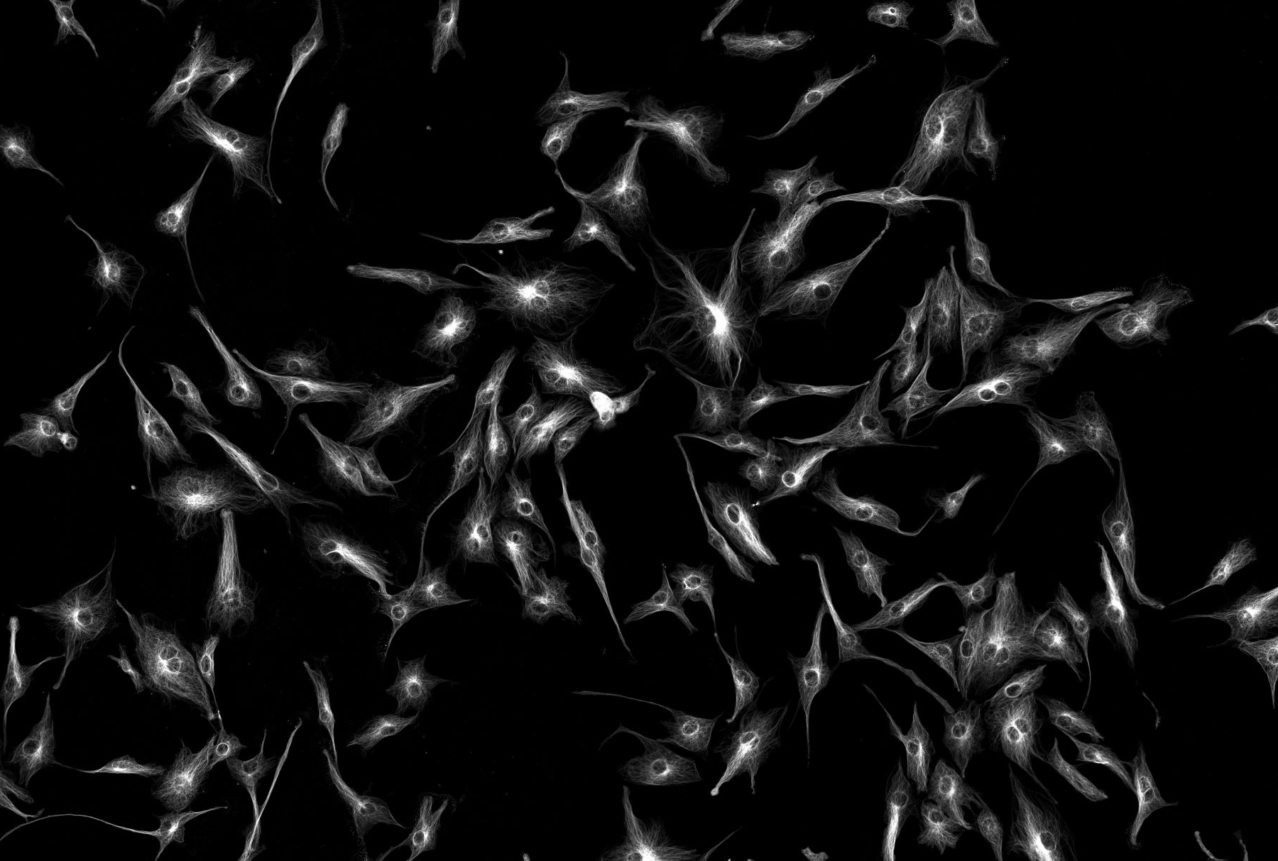
This instrument is based around an inverted Leica SP8X white light confocal microscope equipped with 405nm and a pulsed White Light Laser (470-670 nm) sources. This white light laser (WLL) source of the Leica SP8X perfectly matches the wavelenght of any fluorophore. Up to eight excitation lines can be used simultaneously. By tuning both excitation and detection, complete excitation-emission spectra can be acquired.With this spectral information, any dye can be optimally excited with minimum cross-excitation and specimen damage.
In addition, the pulsed WLL supplies excitation light for fluorescence lifetime measurements. Each selected wavelength has about 1.5 mW power which can be attenuated with the AOTF (Acusto-Optic Tuneable filters). The WLL is a pulsed laser (80 MHz), allowing time gating of fluorescence life times in combination with Hybrid detectors.
The system has two PMTs for fluorescence, one for transmitted light and two gated hybrid detectors. Hybrid detectors allow Low Dark Noise and High Contrast: Sensitive live specimens need to be imaged under low light conditions in high gain situations. Low noise can become the decisive advantage when it comes to recognizing weakly stained detail. Low dark noise is necessary for maximum signal efficiency, especially when photons are accumulated for more information. Otherwise, noise will pile up in the background of the image.
The Leica HyD offers superior signal-to-noise ratio to help render the finest details from any specimen – even tricky ones, such as highly scattering tissue slices. By reducing dark noise, the Leica HyD automatically improves image contrast. You obtain more information content, and the images are immediately publication-ready without any need for image processing. An incubation box allows temperature and Co2 control. Adaptive focus control (AFC) maintains the focus during prolonged live cell experiments.
Specification
Objectives
Magnification |
NA |
Coverslip |
Other modes |
Immersion |
|---|---|---|---|---|
| 10 | 0.04 | 0.17 | Air | |
| 20 | 0.75 | 0.17 | DIC | Air |
| 40 | 1.3 | 0.17 | DIC | Oil |
| 63 | 1.2 | 0.17 | Water | |
| 63 | 1.4 | 0.17 | Oil | |
| 100 | 1.4 | 0.17 | STED White | Oil |
Filter (epi)
Cube |
Example fluorescence |
Excitation |
Dichroic |
Emission |
Part number |
|---|---|---|---|---|---|
| Leica DAPI | DAPI, Hoechst | 340-380 | 400 | 420-(LP) | |
| Leica FITC | GFP, FITC, AF488 | 450-490 | 510 | 515 (LP) | |
| Leica RHOD | mCherry, AF594 | 515=560 | 580 | 590 (LP) |
Light source (epi)
Broad spectrum fluorescent light
Light source (laser lines)
White Light Laser - any wavelength between 470nm to 670nm
Detectors (cameras or PMTs)
2 HyD, 2 PMT, 1 TL