Nikon A1R
Modalities: invert, confocal, live cell, high content
Overview
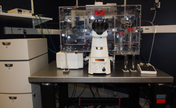
The Nikon A1R is a point scanning confocal microscope based around a fully motorized, inverted Nikon Ti body with hardware-enabled focus correction.
The Nikon A1R has three photomultipliers for fluorescence imaging, one for transmitted light detection and a dedicated photomultiplier array for spectral profiling or if required, unmixing of fluorescently ‘complex’ specimens.
Incorporated into the confocal scan-head are both resonant (fast scanning) (HD 1024x1024) and non-resonant (conventional scanning) scanning mirrors. This allows experimenters to have the option of imaging in high resolution (up to 4096x4096 pixels), at high speed (420fps at 512x32 pixels) or a combination of both.
This arrangement is ideal for live-cell photo-activation and photo-bleaching studies where resonant scanning can be used to capture images at high-speed and non-resonant scanning can be used to target a region of interest for photo-activation.
Resonant scanning mode has added benefits; for live-cell experiments, photo-sensitive specimens remain live for longer and fluorophore bleaching is significantly reduced. The system will also perform regular 2D, 3D and 4D imaging.
The microscope is fully-enclosed in a heated environmental chamber and has the ability to set, maintain and monitor temperature, humidity, CO2 and O2 levels as imaging conditions dictate.
The system is run through Nikon’s versatile Elements software package and incorporates wizards to perform common imaging tasks such as FRAP, FRET and colocalisation as well as data quantification.
Imaging modes
- Multi point x,y (2D) with tiling
- Volume x,y,z (3D)
- Time lapse x,y,z,t (4D)
- FRET
- FRAP
- Spectral
- Live Cell
Specification
Here you can find the specifications for this microscope.
Objectives
Magnification |
NA |
Coverslip |
Other modes |
Immersion |
|---|---|---|---|---|
| 4 | 0.2 | Air | ||
| 10 | 0.45 | 0.17 | DIC | Air |
| 20 | 0.75 | 0.17 | DIC | Air |
| 40 | 0.95 | 0.17 | DIC | Air |
| 40 | 1.3 | 0.17 | Oil |
Filters (epi)
Cube |
Example fluorophores |
Exctitation |
Dicroich |
Emission |
Part Number |
|---|---|---|---|---|---|
| Nikon DAPI | DAPI, Hoechst | 340-380 | 400 | 435-485 | |
| Nikon FITC | GFP, FITC, AF488 | 465-496 | 505 | 515-555 | |
| Nikon G2A | mCherry, AF594 | 510-560 | 565 | 590 (LP) |
Light source (epi)
Intenslight (Broad spectrum fluorescent light)
Laser lines
405nm, 488nm, 561nm, 647nm
Detectors (cameras, PMTs)
2 x PMT, 2 x GaAsP, 1 x PMT for transmission