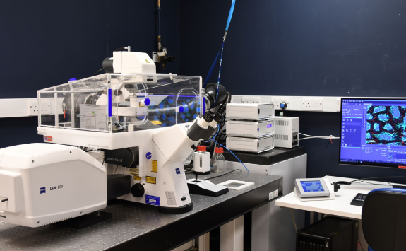
Zeiss LSM800
Modalities: invert, confocal, live cell, airyscan
Overview
The microscope employs software and hardware autofocus correction and is fully-enclosed in a heated environmental chamber with the ability to set, maintain and monitor temperature, humidity, O2 and CO2 levels as imaging conditions dictate.
The system is run through Zeiss’ Zen software package.
Imaging modes
- Multipoint x,y with tiling (2D)
- Volume x,y,z (3D)
- Fluorescence
- DIC
- Time-lapse x,y,z,t (4D)
- Z-stack and multi-color imaging of cell cultures, living sections, organs.
- Imaging + electrophysiology combining mode
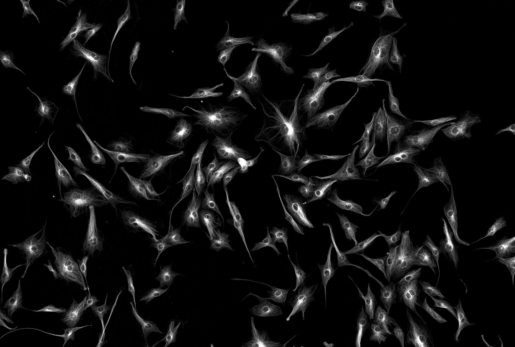
- SuperResolution (AiryScan)
Specification
Here you can find the specifications for this microscope.
Objectives
Magnification |
NA |
Coverslip |
Other modes |
Immersion |
|---|---|---|---|---|
| 5 | 0.16 | Air | ||
| 10 | 0.3 | 0.17 | Air | |
| 10 | 0.45 | 0.17 | Air | |
| 20 | 0.7 | 0.17 | DIC | Air |
| 20 | 0.8 | Air | ||
| 25 | 0.8 | 0.17 | Multi | |
| 40 | 1.3 | 0.17 | Oil | |
| 63 | 1.4 | 0.17 | DIC | Oil |
| 100 | 1.4 | 0.17 | Oil |
Filters (epi)
Cube |
Example fluorophores |
Excitation |
Dichroic |
Emission |
Part number |
|---|---|---|---|---|---|
| Zeiss 49 | DAPI, Hoechst | 335-385 | 395 | 420-470 | |
|
Zeiss 38 HE |
GFP, FITC, AF488 | 450-490 | 495 | 500-550 | |
| Zeiss 43 HE | CY5, AF647 | 625-645 | 660 | 675-715 | |
Light source (epi)
HXP120 - Broad spectrum fluorescent light
Laser lines
405nm, 488nm, 561nm, 640nm
Detectors and Cameras
GaAsP and AiryScan