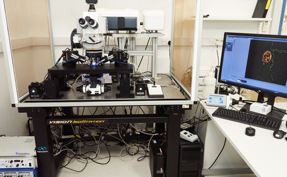
Zeiss LSM880 upright
Modalities: upright, confocal, electrophysiology, airyscan, UV
Overview
The LSM880 with Electrophysiology set-up is a point scanning confocal microscope.
This equipment was funded by [Wellcome Trust, 2017], please reference the funder and [208339/Z/17/Z], in your publication.
This system is built on an upright microscope body-Examiner Z1. It is a semi-automated microscope with a stationary fixed stage.
The objectives are specifically selected for electrophysiology therefore the majority of them are water immersion objectives.
The microscope is set-up for electrophysiology with the following set-up:
- Faraday cage
- 2 micromanipulators
- Axoclamp 900A
- Digidata 1550B
- Temp. Control
- Humbug
Specification
Here you can find the specifications for this microscope.
Objectives
Magnification |
NA |
Coverslip |
Other modes |
Immersion |
|---|---|---|---|---|
Currently in use: | ||||
| 5 | 0.16 | Air | ||
| 10 | 0.45 | 0.17 | DIC | Water Dipping |
| 40 | 1 | 0.17 | DIC | Water Dipping |
| 63 | 0.9 | 0.17 | DIC | Water Dipping |
Additional available lenses: | ||||
| 2.5 | 0.12 | Air | ||
| 10 | 0.3 | 0.17 | Water Dipping | |
| 40 | 0.5 | 0.17 | Water Dipping | |
| 40 | 0.75 | 0.17 | Oil | |
| 63 | 1.4 | 0.17 | Water Dipping |
Filters (epi)
Cube |
Example fluorophores |
Excitation |
Dichroic |
Emission |
Part number |
|---|---|---|---|---|---|
| Zeiss 90 HE LED | DAPI | 370-400 | 405 | 410-440 | |
| FITC | 450-488 | 493 | 499-529 | ||
| AF5686 | 540-570 | 575 | 580-604 | ||
| AF647 | 615-643 | 653 | 659-759 | ||
|
Zeiss 38 HE |
GFP, FITC, AF488 | 450-490 | 495 | 500-550 | |
| Zeiss 63 HE | AF568, mCherry, TRITC | 560-584 | 590 | 598-660 |
Light source (epi)
Colibri’ 7- LED fluorescent light (385/30, 423/44, 469/38, 511/44, 555/30, 590/27, 631/333)
Laser lines
355nm, 458nm, 488nm, 514nm, 561nm, 594nm, 633nm
Detectors and Cameras
Hamamatsu ORCA-Flash-4.0 V3 (Monochrome for fluorescence imaging) GaAsP and AiryScan