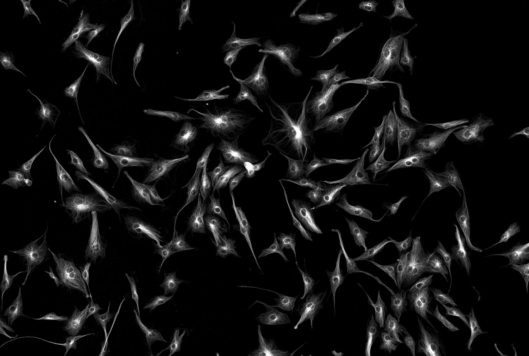
Light-Sheet Microscopy
Fluorescence lightsheet microscopy excites the sample with a thin plane of light and then captures fluorescence emission perpendicular to that. It offers some benefit by only exciting the plane being imaged, in contrast to conventional confocal or widefield fluorescence techniques which can potentially bleach the sample in out of focus planes. Also, the plane can be thin enough to provide a level of confocality, but with the benefit or speed, since the entire plane can be imaged at once using a camera. Combining a sensitive camera with fast movement of the excitation light (or the sample), allows very rapid 3D imaging. This can be achieved with reasonably low excitation light levels (so ideal for live imaging), although can suffer from scattering problems if the sample isn’t transparent enough. Common uses are for zebrafish embryos and tissue samples/ organisms which have been cleared in a way that is compatible with the imaging optics.
- Find out more: PACT
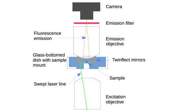
The Leica Digital Light sheet (DLS) available in the Bioimaging Unit utilises the same concept, except that instead of creating a plane of light it uses a standard confocal scanner unit to sweep a confocal point of light across the sample very quickly to generate a virtual sheet. Combining this with the integration time of the camera produces an evenly illuminated sample. It is configured around a standard confocal for the excitation and uses two 45o mirrors around an emission objective to allow generation of a perpendicular ‘plane’ of light through the sample (Figure 1), which is mounted in a glass bottomed dish slightly raised to allow positioning of the mirrors on either side. Two mirrors allows excitation from either side, helping overcome the problem of scattering through thicker/ more opaque samples by combining the results from both. Typically, the DLS captures a single plane in one colour every 50ms and can accommodate samples up to 3-4 mm wide.