QUAREP
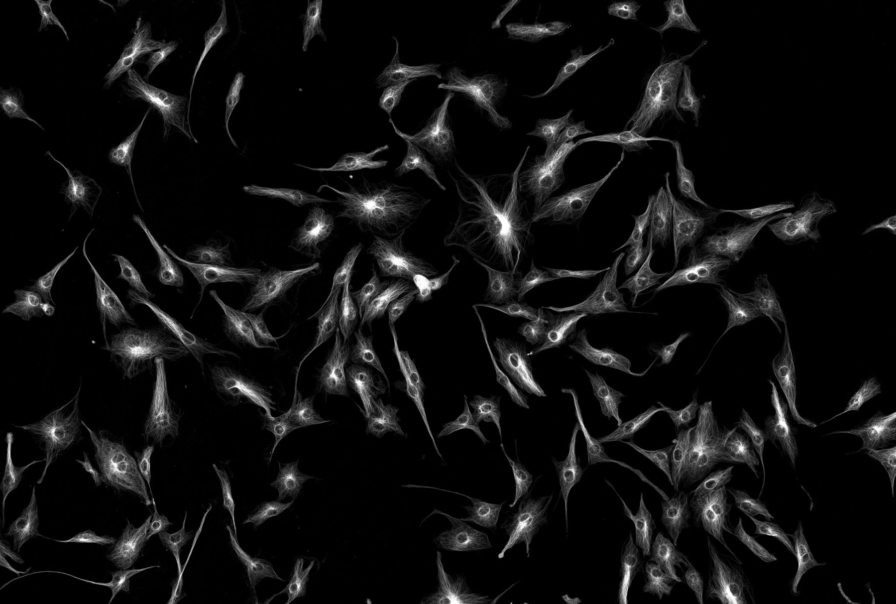
Since their introduction and widespread use in the late 17th Century, microscopes have moved from qualitative, image-collecting tools to automated, complex instruments capable of acquiring information-rich images. Indeed, microscopy now underpins a lot of biomedical research and is being used to provide the spatial context to many emerging ‘omics’ techniques. Observing a sample is often insufficient; there is a need to back-up these observations with quantitative information answer often complex biomedical questions. The quality of these observations and data are only as good as the quality of the microscope used to make them: thus, it is important that microscope ‘quality’ is understood, documented and made available with the image. With this information to hand, researchers and the public will have full confidence in the imaging data collected, interpreted and published from such instruments.
The problem is that standardisation for fluorescence microscopy is not widely practised in the scientific community: a recent large (from approximately 200 imaging labs) global microscopy-user community survey highlighted the inconsistencies when it comes to choosing which microscope Quality Control (QC) metrics to record and how frequently they are performed. Alarmingly some microscopes used in publications receive little in the way of quality control.
Paradoxically, enforced virtual communication methods over the pandemic have brought together communities, and this has benefitted the formation of QUAREP-LiMi (QUality Assessment and REProducibility for Instruments & Images in Light Microscopy, www.quarep.org). Members of the BioImaging facility team have played an important role in this. QUAREP-LiMi brings together over 270 microscopists; researchers; manufacturers; national and international standard-setting organisations and scientific publishers. The initial aim is to gain consensus and identify a set of QC tests that should be performed on microscopes at defined intervals. Work will then lead onto standardising QC samples and automating QC data capture and analysis. This will, we hope, ‘demystify’ the subject and encourage good practise for microscope maintenance and promote standardisation between labs and research facilities employing light microscopy.
Alex Laude / Glyn Nelson / Veronika Boczonadi
Nature Method published two stories - 27/05/2022
Two stories were published online in the Technology Features section of Nature Methods about the work in QUAREP-LiMi:
- Imaging standards to ease reproducibility and the everyday
- The making of microscope camera standards
They will be printed in the July edition of Nature Methods.
Illumination Power and Illumination Stability
Objectives
Comparison of fluorescence intensities between images requires measurements of the illumination power and stability of the excitation light source. WG1 aims at establishing a recommended protocol for measuring the stability of a light source during both short- and long-term image acquisition sessions using calibrated external power sensors. This initial aim will be extended later to measure the absolute flux of light through the illumination path and irradiance of the sample. The initial protocol will be designed around lasers on confocal microscopy platforms (raster scanning and spinning disks). It will be later modified towards other microscopy techniques (widefield, TIRF, light-sheet, super-resolution).
Publication
See our protocol for Illumination Power and Illumination Stability: Protocol
Further information can be found on the QUAREP website.
Detection System Performance
Objectives
WG2 focuses on the detection system, comprising the detection path and its detector(s), and how it measures the signal from the sample.
Members of WG2 aim to standardize the characterization of the detection system performance and create standard procedures for monitoring it over time, thereby revealing performance issues that could affect data reproducibility. Therefore, WG2 will define universal, externally measurable parameters applicable to any type of detector (e.g., photons, linearity, noise), together with measurement tools and protocols for measuring these parameters from common detector types. These universal parameters will be specified according to each distinct type of detector’s internal parameters, which have already been defined by the community. They will enable the evaluation and comparison of different detection systems, thus pinpointing the most suitable technology for given applications.
Further information can be found on the QUAREP website.
Uniformity of Illumination Field - Flatness
Objectives
Illumination field uniformity is critical for quantitative imaging when comparing fluorescence intensities across a field-of-view (FOV) or a large tile of images capturing an entire sample. If the illumination is not constant over a large area, the fluorescence intensities will not represent the inherent fluorescence but rather the location within the image. Thus, WG3 aims at defining a set of universal protocols to assess the uniformity of illumination (i.e., “flatness” of field) over the FOV of any photon-based imaging system and allow for correction of any non-uniformity. These protocols will identify the necessary tools, the procedures required to perform the measurements, and the analysis methods required for their interpretation. WG3 will also define criteria regarding the cut-off for acceptability and the need for correction. A database will be created with ideal images of uniform fields-of-view from different microscope modalities and settings to be used as a reference by the community and validate the protocol and criteria.
Further information can be found on the QUAREP website.
System Chromatic Aberration and Co-Registration
Objectives
Chromatic aberration refers to possible artifacts caused by the wavelength dependency of an imaging system’s optical properties, with the result that two colors arising from the same physical location within the sample appear separated in the image. Such artifacts result from the optical design of the system (e.g., well-corrected versus poorly corrected objective lenses), the manufacturing tolerances of the system components, and the alignment of the optical components.
Co-registration accuracy more generally refers to the system’s ability to co-localize dyes of different wavelengths emitting from the same object within a particular experimental set-up. This can be affected by both the experimental set-up and the system architecture. Working within the assumption that microscope users are ultimately interested in co-registration accuracy, WG4 aims to use sub-resolution and larger multi-colored bead preparations to measure co-registration accuracy. Alternative tools for performing these measurements will also be evaluated. WG4 will compare reproducibility across different laboratories to determine the best protocol.
Further information can be found on the QUAREP website.
Lateral and Axial Resolution
Objectives
This WG focuses on the microscope lateral and axial resolution, which is essential for reporting size measurements of near- resolution limit objects or distances between them. Resolution is highly related to the objective quality but depends strongly upon other parameters ranging from the sample preparation to the signal detection.
The WG aims to define sample preparation, image acquisition, and data analysis protocols for testing resolution, first using sub-resolution fluorescent bead preparations and second employing alternative pattern-based methods. Monitoring the resolution (Point Spread Function in the case of beads) over time will identify possible aberrations in the system. Pooling the data from multiple laboratories within the WG will allow them to compare reproducibility for sample preparation, data acquisition, and data analysis tools, thereby determining a robust, easy-to-use protocol to propose to the community.
See our protocol for Lateral and Axial Resolution: Protocol
Further information can be found on the QUAREP website.
Stage and Focus - precision and other
Objectives
The mission of WG6 is to ensure the performance and QC of stage platforms and sample holders and the optomechanical focus of the optical system as it relates to X, Y, Z movement, stability, reproducibility, and repeatability. The goals are defining the terms typically used to address QC, providing standardization of the measurements and testing protocols, and establishing performance benchmarking levels. Though initially applying these towards confocal light microscopy, the WG will endeavor to include details for standard incident light fluorescence microscopy and more advanced techniques such as super-resolution and light-sheet microscopy.
See our protocol for Stage and Focus: Protocol
Further information can be found on the QUAREP website.
Microscopy Data Provenance and QC Metadata
Objectives
The Work Group 7 focuses on various aspects of Image Metadata (initially primarily Microscopy Metadata but also Sample and Analysis Metadata). These span from defining and continually revising the 4DN-BINA-OME (NBO) Microscopy Metadata model, to facilitating metadata capture through user-friendly tools, to integrating the captured metadata into cloud-ready storage containers such as the newly developed Open Microscopy Environment (OME) Next-Generation File Format (NGFF). To promote the uptake of metadata tools and models, educational material and outreach strategies are also developed.
To carry on these tasks, WG7 is organized into four Subgroups (see “Subgroup” tab below), each of which carries out specific objectives. These subgroups are created, paused, or dissolved on a needs basis as agreed by the WG7 as a whole.
The general meeting serves as a space for reporting back on the progress that has been achieved within the Subgroups, connecting the dots, strategic planning, and general discussions.
The Subgroups (see “Subgroup” tab below) identify specific tasks to be carried out to move our work forward. To participate, please first fill in the general QUAREP-LiMi membership form and then enter your name here. Still confused? No worries, we have a specific list of tasks that you can check out to know exactly how you can lend a hand.
Further information can be found on the QUAREP website.
Education, training and outreach
Objectives
The remit of WG8 is to relay both short and long-term goals of QUAREP-LiMi by the publication of a set of White Papers to communicate and seek cooperation from the community.
The principal aim of these White Papers is to promote QUAREP-LiMi to:
- prospective new members: to actively engage with the work of QUAREP-LiMi
- imaging scientists and bioimage analysts: to raise awareness of QC issues
- group Leaders/Principal Investigators: to engage a critical mass of academic researchers (top-down)
- research scientists (graduate students and postdoctoral researchers) with expertise in the specialized WG topics and imaging scientists: to influence the research group leaders (bottom-up)
- scientific publishers: to raise the quality of methods reporting and rigor and reproducibility in publications
- leads (CEO/directors) of companies and commercial application specialists: to work alongside QUAREP-LiMi to facilitate ease of measurements and reporting
- prospective funders (funding agencies, private sponsors): to support the work of this initiative
Further information can be found on the QUAREP website.
Overall Planning and Funding
Objectives
The principal aim of WG9 is to coordinate and promote the activities of QUAREP-LiMi. Within this WG, there is representation from all other WGs in addition to key global, regional, and national microscopy communities. WG9 will also liaise directly with corporate partners, scientific publishers, and funding bodies.
WG9 will focus on the following activities:
- ensure that the output of QUAREP-LiMi achieves maximum impact within the imaging community by raising awareness of the need for QC across all stakeholders in light microscopy (via white paper, website, publications)
- seek to obtain support from our corporate partners (microscope manufacturers/technology companies)
- obtain funding and support from national bodies, scientific publishers and learned societies to help cover the activities of QUAREP-LiMi (allow us to stage physical meetings, cover publication costs, help with organization and add impact)
- keep stakeholders informed and share information through a regularly updated website and tools database (towards internal and external communication and impact)
- coordinate QUAREP-LiMi WGs and future QUAREP-LiMi meetings (virtual and physical)
Further information can be found on the QUAREP website.
Image Quality
Objectives
Good image quality (IQ) is essential for any subsequent image processing, analysis, and presentation steps. However, the notion of IQ is very broad and encompasses concepts that might differ between various microscope types. The aims of WG10 are 1) to define a set of basic IQ parameters (quantitative criteria, metadata, QC metrics) for light microscopy; 2) to weight the significance of the individual parameters for different experimental techniques and microscope types; and 3) to facilitate the assignment of a microscope- and experiment-specific QC rating to individual images. Ultimately, WG10 will work to summarize the upshot of these steps in the form of easy-to-use workflows. The integration of IQ ratings as part of the metadata associated with every imaging dataset is a long-term goal of this WG.
Further information can be found on the QUAREP website.
Microscopy Publication Standards
Objectives
WG11 will work together with scientific publishers to promote the adoption of best practices in the reporting of metadata (for both image acquisition and analysis) throughout scientific journals and books. Only by ensuring all relevant constituents researchers and imaging scientists submitting publications and designing research; editors, scientific publishers, and reviewers monitoring and preparing publications; and funders, researchers, and educators evaluating and disseminating publications) are working in concert can we raise the bar to ensure reproducibility in imaging experiments. WG11 will focus on the following activities: 1) inform scientific publishers of the standards and metadata put forward by the other QUAREP-LiMi Working Groups; 2) liaise with and encourage individual journals to modify their imaging guidelines to align with these recommendations; 3) work together with the scientific publishers to enforce high standards of imaging metadata reporting in all research works accepted for publication; 4) facilitate the involvement of technical reviewers with significant microscopy expertise during the review of papers that rely heavily on imaging techniques; 5) work together with publishers to promote and increase the appropriate acknowledgement and co-authorship of imaging scientists and core imaging facilities in publications; 6) encourage publishers to compel authors to make raw imaging data available if, and when, required for validation of published research and to make reasonable suggestions regarding duration of storage of raw imaging data relevant to published results; 7) propose minimum standards for figure quality, figure colour selection, scale bars, inserts, annotations and labelling, in order to render all microscopy figures easily interpretable by experts and non-experts alike.
Further information can be found on the QUAREP website.
Image Visualization and Analysis
Objectives
The Image Analysis and Visualization Working Group (WG 12) will provide guidelines for scientists to publish accessible and understandable images, including their reproducible processing and analysis procedures. Publishing of light microscopy image data should include key information about image acquisition (Link:WG11), image reconstruction, processing and analysis, as well as image visualisation and image data availability (safeguarding e.g. in repositories).
WG12 will provide a white-paper, guidelines, training materials, teaching slides with example images, and an application of the guidelines in a video-tutorial. WG12 has members from academic and corporate research, and publishing. Members are active in image analysis, image publishing, image software design, and data visualisation.
Further information can be found on the QUAREP website.
Phototoxicity
Objectives
In live fluorescence microscopy, phototoxicity describes the phenomenon by which the light used for fluorescence excitation leads to physiological changes in the observed living sample. Fluorescence imaging itself can change the physiology, and the conclusions drawn from such observations can be erroneous and misleading: Phototoxicity may cause subtle changes within the sample that are not easy to notice visually, and researchers may observe a physiological process for the first time, for which “normal” behaviour is not known. The goal of the phototoxicity working group (WG13) is to raise awareness of phototoxicity in the wider imaging community and share the best ways to assess, minimise and report it. Key aims of WG13 are:
- to bring together the international microscopy community to raise awareness about the issue of phototoxicity in live fluorescence microscopy. This includes imaging scientists working in core facilities, researchers and trainees (graduate students, postdoctoral fellows), scientific publishers, manuscript/grant reviewers, and manufacturers of optical instruments and software
- the WG members will generate content and give phototoxicity workshops and short talks at different scientific meetings in their regional area and in their scientific field (e.g. biophysics, neuroscience…). WG13 will also periodically produce educational material (blog articles, videos, reviews) with up-to-date references (peer-reviewed publications, white papers and industry notes) regarding phototoxicity
- to develop and disseminate general recommendations and best practices on how to measure, mitigate and report phototoxicity. For example, how to produce dose–response curves, how to avoid illumination overhead, and what biological readouts to use
- to establish a consensus on phototoxicity-related terminology. For these points, we intend to work with the Microscopy Publication Standards group (WG11)
- to work with the Illumination Power group (WG1) to encourage researchers to measure incident light power for all live cell experiments
- to collaborate with industry partners for the promotion of hardware settings and techniques that verifiably enable live imaging with low phototoxicity
- to obtain funding that enables quantitative studies, educational activities and community building centered on phototoxicity
Further information can be found on the QUAREP website.