Super-Resolution Microscopy (Nanoscopy)
Every so often a new technique, approach or vision comes along that challenges accepted methods and dogma. One may argue that over the past five years or so HD and now 3D TV has transformed our home viewing experience. In a similar way, super resolution microscopy although being around for a similar if not longer period of time, threatens to shake up light microscopy. Like HD TV, super resolution microscopy offers the potential to visualize structures in greater detail but in its case, the benefits are not brought about simply by filling the image with more pixels. Indeed, the resolving power of a standard compound microscope is limited by the wave-like nature of light such that simply increasing the pixel density of the captured image has little effect on the resolving power of the system. Abbe’s principles dictate that even with ‘perfect’ optics, it is only possible to resolve details half the wavelength of the incident light. In practice, this means that the lateral (X-Y) resolution limit of GFP–labelled structures (488nm excitation) is at best around 250 nm; axial (Z) resolution is approximately 500 nm.
Armed with novel fluorescent probes and innovative methods with which to use and visualize them eg. structured illumination and deconvolution techniques, cell biologists and microscopists are pushing conventional light microscopy to its limits. There is however, a genuine requirement to probe structures, complexes and individual proteins beyond it. Naturally, this is where Electron Microscopy (EM) takes over but not all biological systems are amenable to EM analysis; it practically impossible with EM to image specimens in their unperturbed state and many EM techniques themselves introduce artifacts. Super resolution microscopy promises to extend the resolving power of light microscopy into that of EM and with it allow the observation of cellular processes in a different light.
So should we now disregard Abbe’s principles, has this diffraction limitation been broken? In a nutshell no, although Abbe I’m sure if he were alive would have a wry smile on his face; super resolution microscopy techniques successfully overcome the diffraction limitations by taking advantage of the way in which the incident illumination interacts with the specimen and in some cases, exploit the properties of the fluorescent label itself. Over the past decade or so a variety of ‘super resolution’ methods have been developed. In the facility we employ a number of super-resolution capable microscopes and techniques these are listed below:
Find out more:
- Fluorescence nanoscopy in cell biology (Nature article)
- Super-Resolution Microscopy (Microscopy U article)
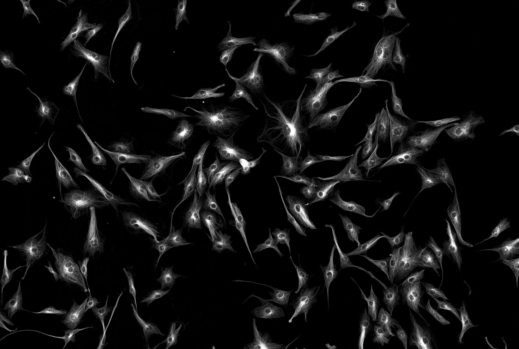
Zeiss AiryScan
The Zeiss Airyscan detector is one of the first significant improvements in optical sectioning in the last 25 years resulting in better resolution and improved signal-to-noise ratios.
Typically, confocal microscopes allow light only from one point to reach the detector using a special kind of aperture, called a pinhole. Light coming from planes above or below the focal plane is out of focus when it reaches the pinhole and those are rejected. Too large a pinhole, then too much light or “noise” gets through; too small a pinhole, then not enough light or “signal” gets detected. By varying the pinhole diameter, the degree of confocality can be adapted to best fit – usually at one Airy Unit (1 AU). However, the Airyscan detector is an array of 32 detector elements and it would use, and not reject the light outside the area of 1 AU. With this novel detector design, where each detector element functions as a single very small pinhole, a 4-8 times improvement in signal-to-noise ratio (SNR) can be achieved as compared to imaging with conventional confocal GaAsP detectors and the resolution can be increased up to 1.7 fold.
Max typical resolution: 150, 150, 350 (nm, x, y, z)
Multicolour: yes
Live cell: yes, ideal due to increase S:N
Special dyes required: No, standard widefield fluoresence and confocal dyes will be suitable
Sample prep: easy no additional preparation requirements over confocal and widefield microscopy we recommend using Prolong Glass (mounting media), high precision coverslips
Further reading:
https://pubmed.ncbi.nlm.nih.gov/34028713/
ZEISS LSM 880 with Airyscan: Beam Path and Principle of Airyscanning - YouTube
STED (STimulated Emission Depletion) microscopy
Technique: Two laser beams are used at the same time (confocal uses only one).
One (A) stimulates the dye to produce diffraction limited fluorescence (~250nm as in confocal), the second one (B), shaped as a dougnut around the first one depletes (blocks emission of fluorescence) the dye everywhere (C) except in the centre (white area). The final detected fluorescence will be A-B. The photons in the area (C) receive both (A) and (B), are depleted and for imaging purposes invisible to the detector.
Increasing the depletion laser the (B) area gets smaller, leading to increased resolution (and also more bleaching and photo-toxicity).
STED images are much dimmer than confocal ones since most of the photons are depleted (lost) but you gain resolution. Post-processing image restoration (deconvolution) greatly helps to collect as much image detail as possible
Max typical resolution: 50, 50, 150 (nm, x, y, z)
Multicolour: yes (max 3). AF594 + ATTO647N + AF532 is the best 3 colour combination
Live cell: yes but not ideal due to photo-toxicity
Special dyes: not really but only a small selection of the standard ones will perform well
Sample prep: Easy when using Prolong Glass (mounting media), high precision coverslips and 2x primary and 2x secondary (compared to confocal amounts). Users should avoid UV counterstains such as DAPI and Hoechst.
Further reading:
STORM (STochastic Optical Reconstruction Microscopy)
Technique: STORM (and other related Single Molecule Localization Microscopy) captures single photons in order to localize molecules in space.
If all the molecules in a sample are illuminated at the same time (as happens normally) they emit fluorescence at the same time. This means that the diffraction limited green spot (min ~250nm) could be produced by (a) several small red molecules or (b) by a big one. However, if we limit the amount of excitation light only a single molecule inside a diffraction limited area is ON at one time you can better localise by repeating the process until you have enough spots to create an image based upon confidence of localisation.
Max typical resolution: 30, 30, 70 (nm, x, y, z)
Multicolour: yes, but better with one.
Live cell: no due to buffer.
Special dyes: yes, ones that blink such as AF647.
Sample prep: needs care and special buffers.
Further reading:
https://www.microscopyu.com/tutorials/stochastic-optical-reconstruction-microscopy-storm-imaging
SIM (Structured Illumination Microscopy)
Technique: a laser-based wide-field microscope technique where a movable diffraction grating is inserted into the excitation beam path. The diffracted laser beams interfere with each other at the focal plane and create illumination in stripes. This stripe patterns if superimposed with the sample generates a so-called Moiré effect.
With standard (homogeneous) illumination, objects separated by a small distance (~250nm), are not visible. Under structured illumination the final super-resolved image is reconstructed after collecting several images at different orientation of the structured illumination.
Max typical resolution: 120, 120, 170 (nm, x, y, z)
Multicolour: Easy
Live cell: yes, with care over light input due to multipleimages of the same plane.
Special dyes: No
Sample prep: Easy
Further reading:
http://zeiss-campus.magnet.fsu.edu/articles/superresolution/supersim.html
https://www.nature.com/articles/s41592-018-0211-z?WT.feed_name=subjects_fluorescence-imaging