Intelligent Sensing and Communications
Developing real world solutions for communications, sensing and signal processing.
The Intelligent Sensing and Communications (ISC) group was first established in 1989.
Our research explores the whole spectrum of communications and digital signal processing. This includes underpinning theories and applications with industry leading partners.
The group's research comprises three themes:
- communications
- signal processing
- sensor systems
We deliver world leading, high impact research in these areas. It supports world-class education through our BEng, MEng, MSc and PhD programmes.
The current and future sensor systems (e.g. sensors in smart devices), will provide ever more data for analysis. The research motivation is multimodal data processing for next generation of artificially intelligent automated systems.
We aim to contribute AI that will enable significant advances towards the vision. The specific aims for healthcare, security and surveillance are:
- intelligent sensing led AI for mental health
- multimodal human behaviour analysis including anomaly detection
- multiple human identification and tracking in cluttered and congested environments
- multimodal speech enhancement and separation in challenging environments
Intelligent sensing led AI for mental health
Intelligent sensing involves:
- audio
- video
- touch/keypad
- text
It captures attention deficit hyperactivity disorder (ADHD) health symptoms.
Trustworthy AI helps clinicians to perform early ADHD diagnosis. This enables the administering of effective treatment.
Multimodal human behaviour analysis
Human anomaly detection is a challenging task for reliable artificial intelligence (AI). The behaviour of humans requires robust features for modelling, indexing and classification.
This research is on:
- multimodal action recognition
- contextual information retrieval
- information fusion for human behaviour analysis
We research advanced deep learning techniques. The main focus is to provide network explainability and human informed system.
Multimodal human identification and tracking
This research involves core signal processing for multiple human localisation and tracking (MHT) in cluttered and congested environments.
The core signal processing techniques providing the solution for MHT include:
- social interaction modelling
- probabilistic clustering
- non-linear filtering
- efficient data association
- stochastic differential equations based particle flow
- multi-level cooperative fusion
Multimodal speech processing
We have made major breakthroughs in establishing complete mathematical proofs for nonlinear signal processing theory. This has enabled us to establish non-linear models for optimal signal separation and information retrieval.
One of our successes is the development of a complete framework using statistical methodologies for:
- signal fusion
- separation of complex nonlinearly mixed signals
- informational retrieval
The work has contributed to the fundamentals of nonlinear signal processing theory. It has also exploited advanced machine learning techniques to pave the way for the research community.
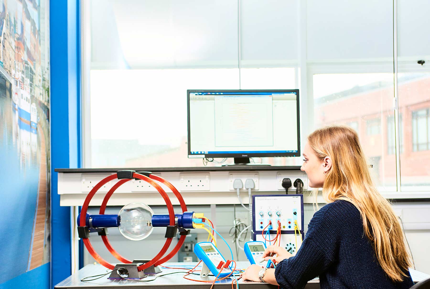
Intelligent sensing laboratory: research facility
Our unique and state-of-the-art intelligent sensing facility is industry-leading in the UK. We built and equipped the Intelligent Sensing Laboratory.
This was built with £0.75M funding from Newcastle University’s £30M Research Investment Fund. The laboratory was opened in October 2016.
The lab is used for cross-disciplinary work. We focus primarily on wearable and non-wearable multi-sensor data acquisition and interpretation for healthcare and security applications.
Research led teaching, supervision and mentoring
Our world-leading research informs our teaching, supervision, and mentoring.
We produce highly employable, multi-skilled graduates and postgraduates. Our graduates are very successful and work all over the globe.
We also supervise and teach talented students to the highest standards through our PhD, MSc, MEng and BEng programmes.
Communications
Our pioneering research enables communication through air, space and the sea.
We design powerful receiver structures and error correction codes for reliable communication in hostile channels.
Our leading-edge underwater communication technologies are used throughout the world, including with:
- divers
- underwater vehicles
- subsea sensors
We use our MIMO base station to carry out research in 5G and future 6G cellular systems. We're developing innovative, scalable approaches to encryption and network security.
Wireless communications
MIMO is a method for sending and receiving more than one data signal at the same time over the same channel, using multipath propagation.
Our research focuses on the design of novel:
- spectrally efficient modulations
- physical-layer waveforms
- error-correction and detection schemes
We work with academia and industry, including:
- Alcatel Lucent
- BP
- Silicon Infusion Ltd
- Cambridge University
- University of Southampton
- Lancaster University
- Defence Science and Technology Laboratory (DSTL), an executive agency of the Ministry of Defence (MoD)
Our facilities
The results of the new algorithms that we develop are supported and verified by practical implementations and measurements. Our excellent research facilities include:
- a massive MIMO base station that enables research in 5G cellular systems and beyond
- an anechoic RF chamber that allows for interference-free measurements
Underwater acoustic communications
Radio waves are severely attenuated by sea water. Thus, sound waves are more effective for underwater communication.
The underwater acoustic channel is affected by severe multipath distortion, Doppler effects and non-Gaussian noise. We are developing advanced modulation, coding and receiver structures to overcome these issues. This enables reliable data transfer through the oceans, over distances from a few hundred metres to tens of kilometres.
Our research includes:
- adaptive equalisation and beamforming for high data rate communication (> 10 kbits/s)
- ultra-reliable low data rate spread spectrum communications (< 1 kbits/s)
- low power, environmentally friendly communication
- underwater tracking and navigation by acoustic ranging and direction-finding
- underwater wireless sensor networks
Our research is supported by EPSRC, NERC, Innovate UK and European Union funding. We collaborate with many other leading institutions, including:
- University of York
- Heriot-Watt University
- National Oceanography Centre
- University of Washington
- University of Zagreb
- University of Rome Sapienza
- INESC-TEC ( Institute for Systems and Computer Engineering, Technology and Science)
DSTL (Defence Science and Technology Laboratory)
Encryption and secure communications
Computer networks and internet use have grown alongside big advances in communications technology.
Threats of interception are increasing in frequency and sophistication. Algorithms to prevent interception and enhance data security are now of primary importance
These algorithms must provide efficient encryption. We provide novel approaches to encryption and protocols, combined encryption and error control. We are developing scalable, secure and robust wired and wireless communications networks.
Sensor systems
Our research focuses on the design and development of:
- novel sensors
- sensor arrays
- non-destructive testing and evaluation
- structural health monitoring
- Internet of Things
We develop hardware, software and signal/image processing algorithms for applications in:
- railways
- pipelines
- aerospace
- marine environment and infrastructure
- medicine, including non-invasive diagnostics and healthcare
Non-destructive testing
We have extensive experience in the field of electromagnetic non-destructive testing and evaluation.
We work across all aspects of electromagnetic NDE, investigating defect detection and characterisation. We develop techniques to quantify macro- and micro-structural changes in components of:
- railways
- pipelines
- aerospace
- offshore wind turbine blades
Our industry partners include global names like:
- Airbus
- British Steel
- General Electronics
We are a pioneer among NDT research labs in the digital age. We develop accurate, effective, efficient analytical and numerical models.
We investigate multi-physics phenomena with multiple parameters and complexities. We use complex non-destructive testing evaluation conditions and apply state-of-the-art NDT & E techniques.
Subsea Passive Acoustic Monitoring (PAM)
The SEAlab team develops hardware, software and signal processing algorithms. These algorithms autonomously detect underwater acoustic signatures from marine mammals (e.g. dolphins). They also pick up propulsion noise from surface and subsea vehicles.
We specialise in computationally light detection algorithms. They run on very low cost and low power devices (edge computing) for long periods at sea.
These devices report detection data in real time via communications networks, including:
- wireless terrestrial
- satellite
- underwater acoustic
This achieves practical examples of the Internet of Underwater Things (IoUT).
Clicks and whistles
Recent examples of this are live dolphin click (echolocation) and whistle (calls) detectors currently operating in the North Sea. These devices are installed in the North Sea and detect clicks/whistles emitted by animals within a radius of up to 3km of the device. Information on detected click trains or whistles is transmitted back to shore and displayed live on a web interface:
Dolphin click trains are displayed with inter click interval (ICI).
Low ICI values indicating rapid click trains, feeding activity and magnitude. This gives an broad indication of the distance from the detector.
Dolphin whistles are displayed in terms of frequency gradient and magnitude. A time-frequency plot appears for a subset of detected whistles allowing the recurring 'signature' whistles to be identified.
Autonomous PAM devices developed by the team can operate for up to 6 months at sea without maintenance. Only a small battery capacity of 4 x 1.5V akaline C cells is required.
Health monitoring
We are developing the next generation of Structural Heath Monitoring system using RFID sensors.
Our research in Advanced Sensors explores the scientific basis for RFID sensors. We study supporting infrastructure technology for measurement of physical quantities. This involves the fields of:
- advanced chip/chip-less RFID sensors
- printable and flexible sensors
- integration of advanced sensors with wireless sensor networks and Internet of Things
- robotic instrumentation or robot assistant instrumentation
- integration of RFID sensors in harsh environments such as oil and gas pipelines
Ultrasound
We are developing a very inexpensive technology for medical ultrasound scanning. We are drawing upon many years of innovation in sonar signal processing.
Our product has a target price of around £200, if sold in large quantities. This would enable the use of ultrasound imaging in applications or regions of the world where it is currently cost-prohibitive.
Technology overview
Our design philosophy uses the minimum possible hardware in the scanning head. It connects to any available PC or mobile device (via USB or Wifi) to perform signal/image processing and display.
We have used a single transducer element. This is because multichannel phased array transducers would far exceed the target cost. We have applied several innovative approaches from sonar signal processing. This has minimised the cost of electronic circuitry. It has produced high-resolution images from a single fixed focus transducer.
The transducer is scanned back and forth across the skin, either by a motorised mechanism or freehand, to gather echo data. The processing software then estimates the motion of the transducer. It performs the focusing operations to generate an image up to four times per second.
Internet of things
We develop cutting-edge autonomous, intelligent systems.
Our research leads to revolutionary IoT solutions in condition and structural health monitoring. We are at the forefront of sensing, communication and information processing.
We design low cost and low power consumption sensors. We build spectrally efficient, robust and secure communications networks. We develop novel approaches to radio frequency identification (RFID) based passive sensing networks and compressed sensing.
We also explore non-linear defect feature extraction and autonomous multi-sensor multi-modality information fusion. Our research in industrial system architecture enables cloud-based computing and artificial intelligence-aided decision-making.
We design ways to assess the location, size, and microstructure of defects in structures and form further decision makings. We do this by applying non-linear system identification and analysis-based signal processing.
We work with cross-disciplinary research teams in Newcastle, Sheffield and York Universities. We also collaborate with strategic industrial partners.
Research themes
Data, digitisation and AI
Much of our research is related to the theme of Data, digitisation and AI. We are heavily involved with AI development, making advances in sensor systems in a number of fields including health and transport. Our work also covers data analysis and building towards automated data processing.
List of current and recent project work
- Multi-task Learning for GEO-located Multimodal Data, Project Leader(s): Dr Mohsen Naqvi, Project Dates: 1 Dec 2022 to 31 July 2024
- Intelligent Sensing and AI for Mental Health Detection, Project Leader(s): Dr Mohsen Naqvi, Project Dates: 1 Sep 2022 to date
- USMART - smart dust for large scale underwater wireless sensing, Project Leader(s): Jeffrey Neasham, Project Dates: 1 May 2017 to 30 April 2020
- Cooperative backhaul aided next-generation digital subscriber loops, Project Leader(s): Dr Charalampos Tsimenidis, Project Dates: 1 October 2015 to 21 December 2018
- Full-Duplex For Underwater Acoustic Communications, Project Leader(s): Dr Charalampos Tsimenidis, Project Dates: 1 January 2018 to 31 December 2020
- INNOvative monitoring and predictive maintenance solutions on lightweight WAGon (INNOWAG), Project Leader(s): Dr Cristian Ulianov (Mechanical Engineering), Project Dates: from 1st January 2017 to 30th June 2019
- Multimodal information processing for security and surveillance, Project Leader(s): Dr Mohsen Naqvi
- Training Network in Non-Destructive Testing and Structural Health Monitoring of Aircraft Structures (NDTonAIR), Project Leader(s): Prof. Gui Yun Tian, Project Dates: from 1st October 2016 to 31st September 2020
- University Defence Research Collaboration (UDRC), Project Leader(s): Professor Jonathon Chambers, Project Dates: 2013 to 2018
- Accurate blood pressure measurement, Project Leader(s): Prof. A, Murray, Project Dates: from 1st July 2016 to 30th June 2019
Underwater communications
Our pioneering research is in underwater acoustic communication and positioning technology. It is commercialised via four industrial partners:
- Blueprint Subsea
- Tritech
- Succorfish
- WSENSE
An example of this is the Seatrac range of products, manufactured under licence by Blueprint Subsea. They are being used worldwide for communication/navigation of underwater vehicles and divers.
Innovations have enabled vast reduction in cost, energy consumption, sound emission and size. This while maximising data rate and range. This is helping to realise the 'Internet of Underwater Things'.
These are large scale, bio-friendly subsea networks. They deployed for marine environment and security monitoring. This work is also contributing to emerging standards for underwater acoustic communication.
Read more about smart dust for large scale underwater wireless sensing
Intelligent sensing for healthcare
Advances in AI and machine learning have led to new systems that can transform healthcare.
Our research is helping:
- to create multimodal data and trustworthy AI for healthcare applications
- clinicians to perform the early diagnosis and thereby administer effective treatment
- us understand health risks
- us predict disease progress
- to provide health interventions for improved patient outcome
Our research and expertise of signal and image processing are proving invaluable. It has had a huge impact in supporting healthcare professions.
Our ongoing research collaborations are with:
- the Adult ADHD Service at the Cumbria, Northumberland, Tyne and Wear NHS Foundation Trust
- the Ophthalmology Department at the Newcastle upon Tyne Hospitals NHS Foundation Trust
PhD opportunities
The ISC is a diverse, internationally-leading group in research, teaching and engagement with industry. Our Group includes a team of academic, research and teaching staff, and PhD and MSc students.
We often have new vacancies within our team. Check out Newcastle University vacancies for current opportunities.
We actively seek and recruit high-quality research students and several PhD studentships are available for Home and EU prospective students. The University also offers scholarships for the very best international students. Please see our postgraduate funding database for all scholarships, bursaries and studentships available.
Teaching
Our Electrical and Electronic Engineering undergraduate programmes give students a path to becoming professional engineers.
Our Electrical and Electronic Engineering postgraduate taught (MSc) programmes equip students with the advanced skills and expertise to help shape the world.
List of academic collaborators
- Cambridge University
- Imperial College
- Leeds University
- Sheffield University
- Manchester University
- University of Bath
- University of Warwick
- Lancaster University
- Queen's University of Belfast
- University of California, USA
- Sidney University, Australia
- Southwest Jiaotong University China
- Queen Mary University of London
- Queen's University Belfast
- Institute of Molecular Medicine USA
- Regional Hospitals, Technical University of Cartagena
List of industrial collaborators
- Cambridge Broadband Ltd
- BAE
- Tritech International
- HW Communications
- Rinicom Ltd
- Shlumberger
- BT Medical
- Foster Findlay
- QinetiQ
- e-Therapeutics
- Torridon
- Nortel
- Rolls Royce
- Alstom Power
- cross-government departments
- British Energy
- The Welding Institute
- Bioinnovel
- Photon fire
- National Physical Laboratory
- RWE nPower
Contact us
For any queries, please contact the head of the group - Professor Jeff Neasham
Email: jeff.neasham@ncl.ac.uk
Telephone: +44 (0) 191 208 8850
Address: School of Engineering, Room E2.11, Merz Court, Newcastle University, Newcastle upon Tyne NE1 7RU