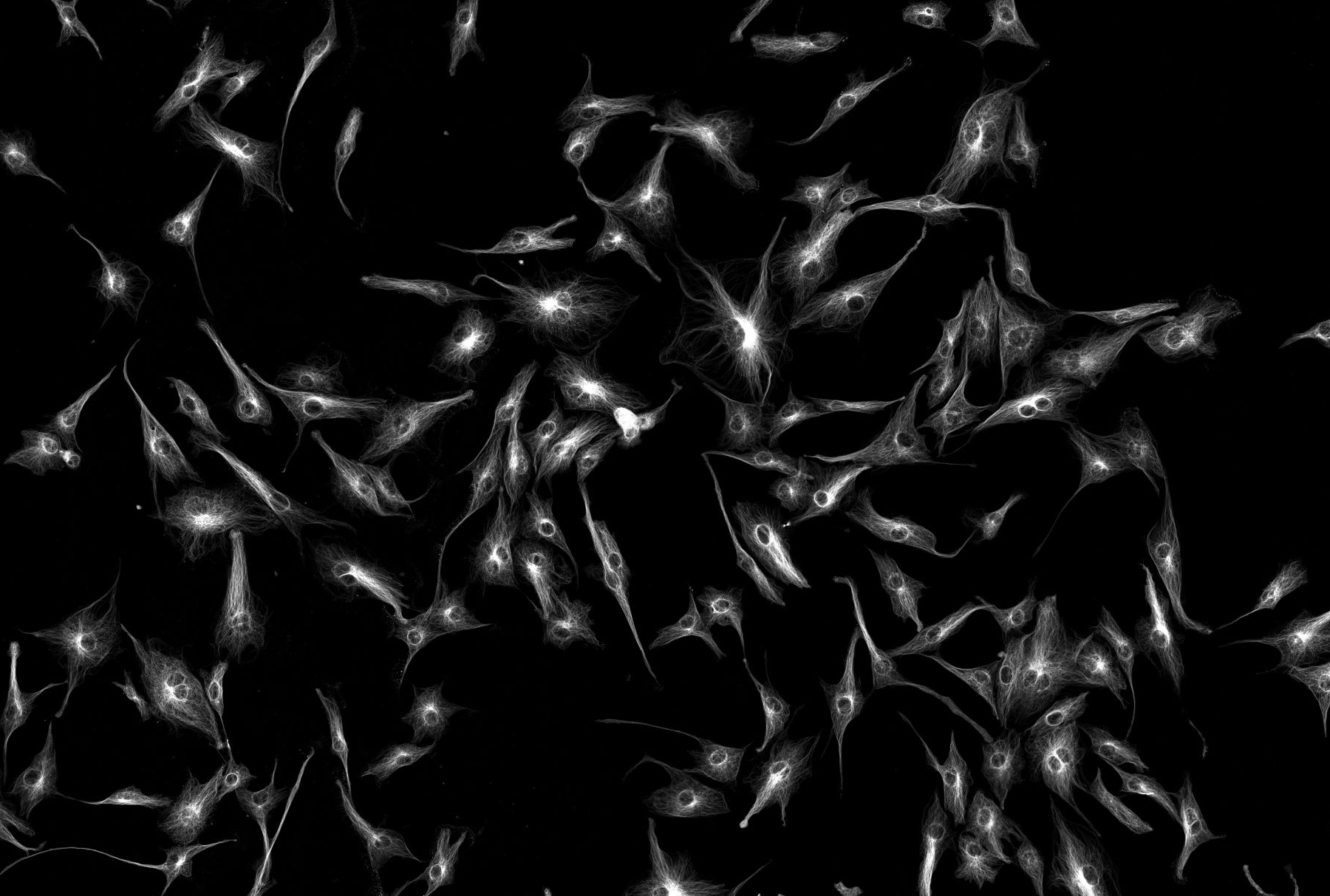
Multiphoton Microscopy
Multiphoton (MP) microscopy, sometimes called Non-Linear Optical (NLO) microscopy typically uses near-infrared (NIR) excitation light which can also excite fluorescent dyes. However, in comparison to standard 1 photon fluorescence confocal microscopy, for each excitation two or more photons of NIR light are absorbed (Figure 1). Using infrared light minimizes scattering in the tissue. Due to the multiphoton absorption, the background signal is strongly suppressed. Both effects lead to an increased penetration depth for this technique, and therefore MP excitation can be a superior alternative to confocal microscopy due to its deeper tissue penetration, efficient light detection, and reduced photobleaching.
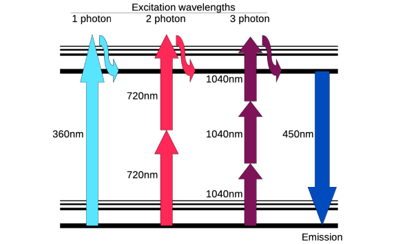
MP microscopy is frequently used for 2 photon excitation, but 3 photon (or more) excitation is possible, and it can also be utilised in other NLO methods, such as Second Harmonic Generation, where two photons absorbed by a suitable material are ‘combined’ to produce one photon with the sum of the energy of the two initial photons. Many biological structures can generate second harmonics, particularly large ordered non-centrosymmetric structures, such as collagen, and can be imaged simultaneously with MP fluorescence.
Find out more: